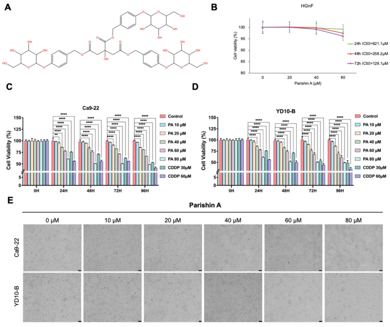
Figure 1.
Parishin A inhibits OSCC cell growth. (A) Chemical structure of Parishin A. (B) Cell viability of normal human gingival fibroblasts (HGnFs) after treatment with 0 μM, 20 μM, 40 μM, and 80 μM Parishin A for 24, 48, and 72 h. (C,D) Cell viability of OSCC cell lines YD-10B and Ca9-22 after treatment with 0 μM, 10 μM, 20 μM, 40 μM, 60 μM, and 80 μM Parishin A for 0, 24, 48, 72, and 96 h, determined by the CCK-8 assay. (E) Morphological changes in YD-10B and Ca9-22 cells after treatment with 0 μM, 10 μM, 20 μM, 40 μM, 60 μM, and 80 μM Parishin A for 0, 24, 48, 72, and 96 h, observed under a light microscope (magnification, 100×). Data are shown as means ± SD from three independent experiments, each with triplicate samples. Asterisks indicate significant inhibition (** p < 0.01, **** p < 0.0001).

