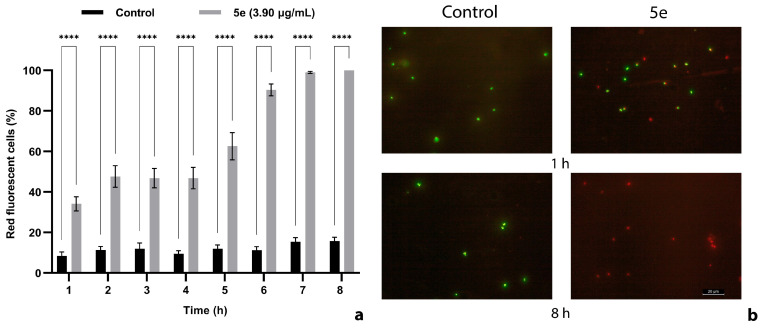
Figure 6.
Effect of 5e exposure on MRSA cell membrane integrity (a). Fluorescent images illustrating the impairment of cell membrane disruption effect, visualized by the uptake of the fluorescent nuclear stain, propidium iodide (b). Exponential-phase cells were incubated for 8 h in PBS supplemented with 5e (final concentration equivalent to MBC) and stained with SYTO 9 and propidium iodide. MRSA cells incubated in PBS supplemented with DMSO served as negative control. Red staining indicates the cellular uptake of propidium iodide due to membrane injuries. Green cells are stained with SYTO 9 indicating intact membranes. Values are the mean of three replicates. Bars indicate SEM. Asterisks denote a significant difference (p < 0.05) vs. Control (**** = p < 0.0001).

