Figure 1.
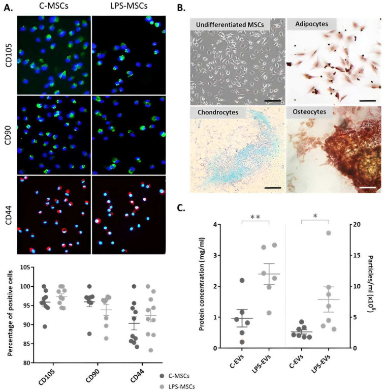
Characterization of C-MSCs and LPS-primed MSCs isolated from rat bone marrow. (A) Immunophenotype of C-MSCs and LPS-MSCs, staining CD105 (FITC, green), CD90 (FITC, green) and CD44 (Texas Red, red) markers. Quantification of the percentage of positive cells for each marker (n = 7–11). The nuclei of the cells were stained with Hoechst (UV light, blue), 20× magnification. Scale bar: 100 µm. (B) Representative images of undifferentiated MSCs and MSCs differentiation towards adipogenic, osteogenic and chondrogenic lineages in vitro. Magnification: 10×. Scale bar: 500 µm. (C) Quantification of the EVs-like particles (right) (n = 5) and protein (left) (n = 7) concentration in C-MSCs and LPS-MSCs media. Data are presented as mean ± SEM (n = 3); * p < 0.05; ** p < 0.01. Abbreviations: extracellular vesicles, EVs.

