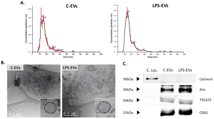
Figure 2.
Characterization of C- and LPS-MSCs-derived EVs. (A) Representative particle size distributions of C-EVs and LPS-EVs’ pools by Nanosight analysis. (B) Representative cryo-TEM images of C-EVs and LPS-EVs, 12kX magnification. Scale bar: 0.5 µm. (C) Detection of Alix, TSG101 and CD81 surface markers in C-EVs and LPS-EVs by Western Blot analysis. Detection of Calnexin only in cell lysate samples as negative control. Abbreviations: cell lysate, C. Lys.; extracellular vesicles, EVs.

