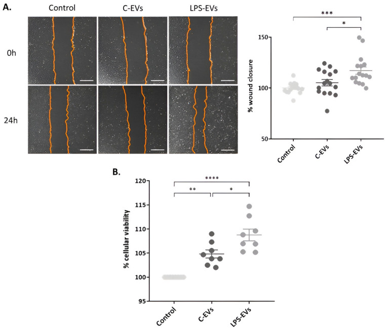
Figure 3.
Effect of MSCs-derived EVs on wound healing and cell proliferation in vitro. (A) Representative optical images of wound healing in HPAEpiC monolayer. Magnification: 20×. Scale bar: 50 µm. Percentage of wound closure in HPAEpiC 24 h after being treated with C-EVs and LPS-EVs. (B) Percentage of cell viability of HPAEpiC 24 h after being treated with C-EVs and LPS-EVs, considering that non-treated cells (control) had 100% cellular viability. Data are presented as mean ± SEM of eight (A) and four (B) independent experiments with two replicates of each condition; * p < 0.05; ** p < 0.01; *** p < 0.001; **** p < 0.0001. Abbreviations: extracellular vesicles, EVs.

