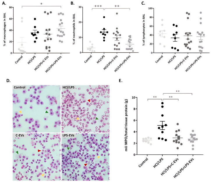
Figure 6.
Effect of MSCs-derived EVs on neutrophil infiltration in the intra-alveolar space in vivo at 72 h. Percentage of (A) monocytes, (B) neutrophils and (C) lymphocytes in BAL by flow cytometry. (D) Representative images of BAL cells cytospins stained with Diff-Quick. Neutrophils (red arrows), macrophages (green arrows) and lymphocytes (yellow arrows) are indicated in each image. Magnification: 20×. (E) MPO activity quantification in lung tissue homogenates (mU/total tissue protein (g)). Data are presented as mean ± SEM (n = 8–14); * p < 0.05; ** p < 0.01; *** p < 0.001.

