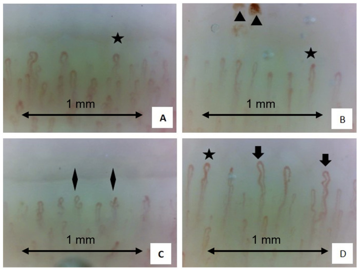
Figure 6.
Examples of four nailfold video-capillaroscopy images recorded among four different sarcoidosis patients (A–D) all in the active stage of disease. (Magnification 200×). Black stars (A–C) indicate the presence crossing capillaries. Black arrows (D) indicate the tortuosity of capillaries. Black triangles (B) show the presence of hemosiderin deposits due to microhemorrhages. Black rhombus (C) indicates the presence of “bushy” capillaries. The capillary count per linear mm results in 7 capillaries/mm (slightly reduced) (Operators: B.R. and L. M., Pulmonology Unit, University of Trieste).

