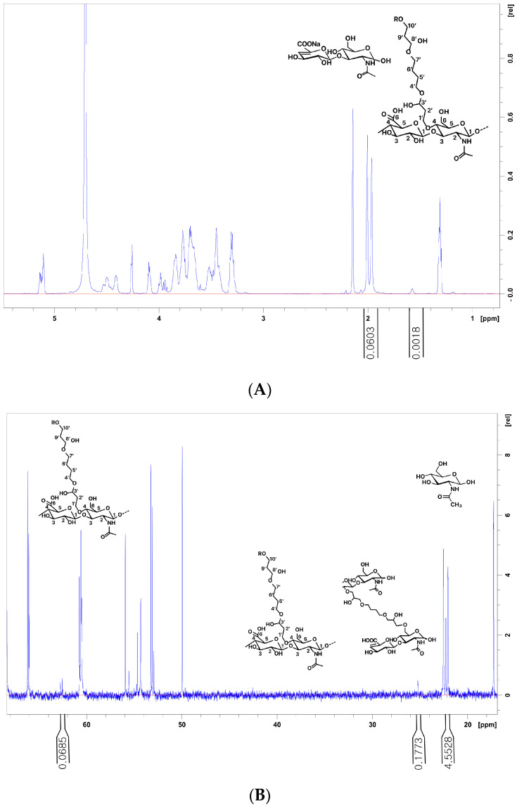
Figure 4.
Different spectra of 1H and 13C nuclear magnetic resonance (NMR) analysis of the hyaluronic acid (HA) filler samples: (A,B) Product 7 has a low MoD but a high CrR value of 0.222 among the tested fillers. (A) The N-acetyl signal (CH3) from HA and BDPE signals (H5′, H6′) used for the determination of the MOD. MoD 1H (%) = (IδH1.7/4)/(IδH2.1/3) × 100. (B) The C10′ signals on mono-linked BDPE are the C5′ and C6′ of both mono- and cross-linked BDPE used to determine the CrR, CrR = 1 − IδC62.7/(IδC25.2/2). The signals and CH3 of N-Acetyl glucosamine are used to determine the MoD, MoD (%) = (IδC25.2/2)/IδC21.9−22.6 100. MoD, degree of modification; CrR, cross-linking ratio.

