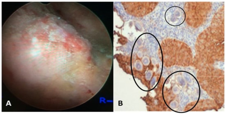
Figure 2.
(A) The presented visual depicts a cystoscopic field of the posterior bladder mucosa exhibiting UGS lesions, granulomas, ulcers, and tumors. (B) Histology from the bladder mass biopsy shows S. haematobium ova (black circles) and Squamous cell carcinoma (SCC) of the bladder (expression of sialyl Lea). The figure is adapted with permission from Vale et al. (2015) and Santos et al. (2021) [37,38].

