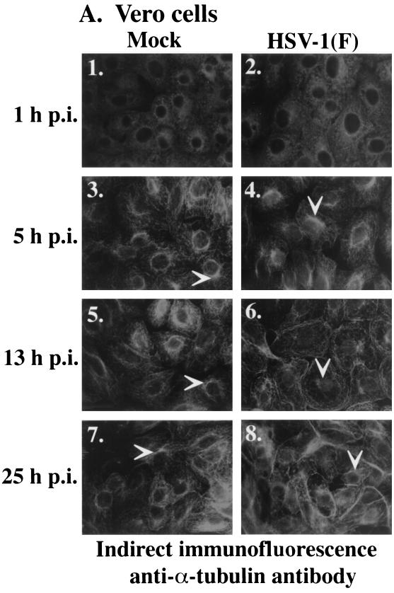
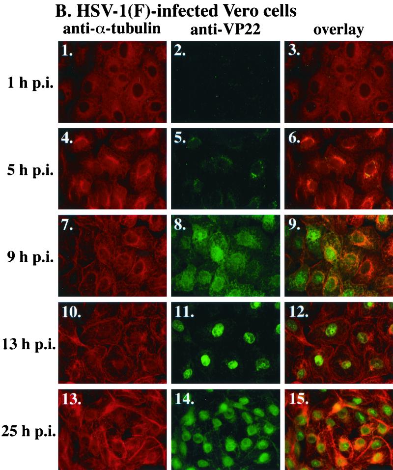
FIG. 1.
Nuclear localization of VP22 during synchronized infection correlates with HSV-1(F)-induced microtubule fragmentation. Vero cells were synchronously infected with HSV-1(F) or mock infected and fixed for indirect immunofluorescence at the times indicated as described previously (37). Cells were stained with antibodies to α-tubulin (A and B) and VP22 (B) as described in Materials and Methods. (A) Gray scale images of mock-infected and infected cells. White arrows mark perinuclear regions (MtOCs). Panels 2, 4, 6, and 8 in A are the same images as panels 1, 5, 10, and 13 in B. Merged images (overlay) are shown in B, panels 3, 6, 9, 12, and 15. All images were acquired under the same conditions.


