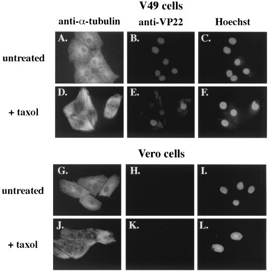
FIG. 3.
Increased detection of cytoplasmic VP22 in taxol-treated V49 cells. VP22-expressing V49 cells (A to F) and Vero cells (G to L), which do not express VP22, were left untreated or treated (+ taxol) with 20 μg of taxol per ml for 20 h. Cells were fixed and stained for indirect immunofluorescence with anti-α-tubulin (A, D, G, and J) and anti-VP22 (B, E, H, and K) antibodies. DNA was stained with Hoechst dye (C, F, I, and L).

