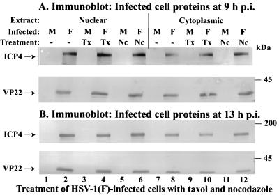
FIG. 5.
Partitioning of VP22 in nuclear and cytoplasmic fractions in cells infected in the presence of taxol and nocodazole. Vero cells were synchronously mock (M) or HSV-1(F) infected in the absence or presence of taxol (Tx) or nocodazole (Nc). At 9 (A) and 13 (B) hpi, nuclear and cytoplasmic fractions (Extract) were prepared, and polypeptides were separated in denaturing gels, transferred to nitrocellulose, and probed with antibodies to VP22 and ICP4 as described in Materials and Methods. Locations of the migrations of molecular mass markers are indicated in the right margin.

