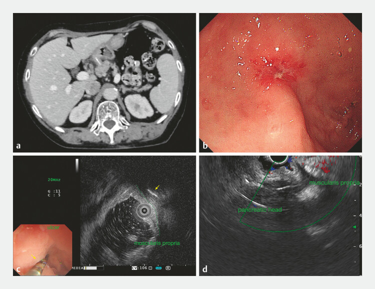
Fig. 1.
Imaging and investigations. a Enhanced computed tomography scan revealed a 3-cm high-density strip penetrating the gastric antrum and extending into the pancreatic head. b Gastroscopy found a small ulcer at the lesser curvature of the antrum. c, d Endoscopic ultrasound revealed the foreign body (yellow arrow) embedded in the muscularis layer near the ulcer and penetrating the pancreas.

