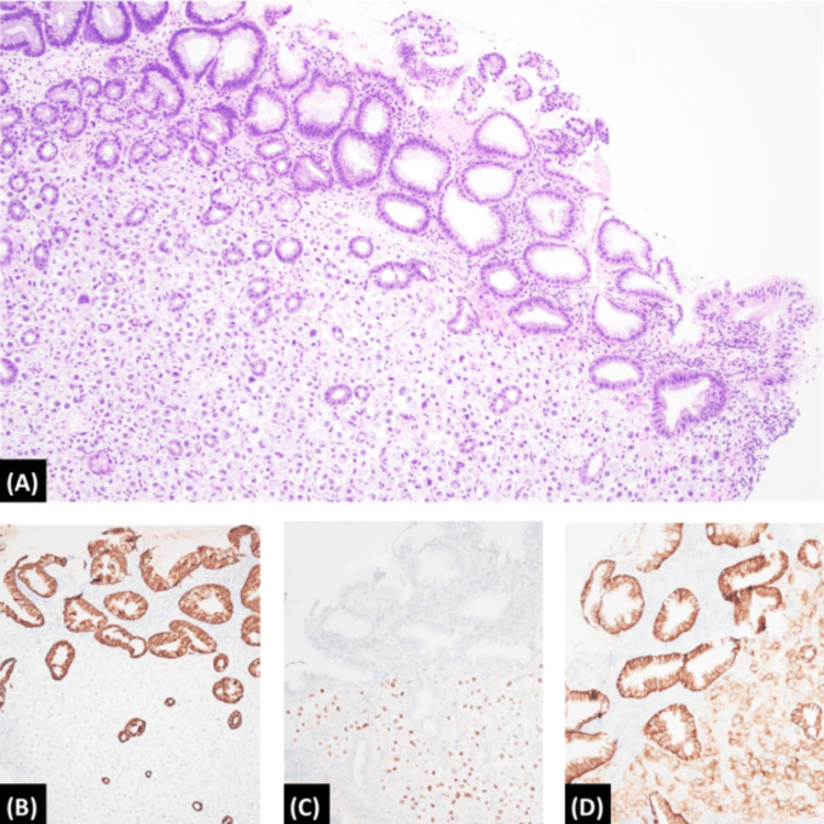
Figure 4. Gastric biopsy histopathology.
Biopsy specimen from the gastric body showed sheets of atypical, epithelioid, and rhabdoid cells with ample eosinophilic to amphophilic cytoplasm and enlarged eccentrically located nuclei, and prominent nucleoli (A; Hematoxylin and Eosin, 100x). By immunohistochemistry, the atypical cells were negative for cytokeratin AE1/AE3 (B; 100x) and positive for both PAX-8 (C; 100x) and carbonic anhydrase IX (D; 100x).

