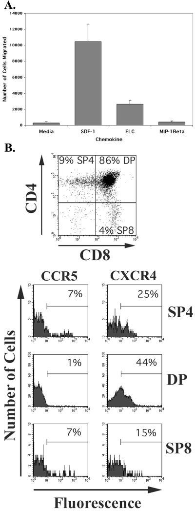
FIG. 7.
(A) Results of a representative chemotaxis assay performed in duplicate with cells derived from pediatric thymus tissue and the indicated chemokines or medium control. The number of cells that migrated to the bottom chamber of the Transwells is shown. The contents of six separate wells were combined for each group and incubated with CD4-FITC and CD8-PerCP, and the light scatter-gated cells were quantified by flow cytometry. Error bars indicate standard errors of the mean of duplicate groups of six cells. (B) Two-color dot plots of CD4 and CD8 expression and single-color histograms of CCR5 and CXCR4 expression on the cells used in the chemotaxis assay shown in A. The CD4 and CD8 stains were used to differentiate the anti-CCR5 and anti-CXCR4 immunofluorescence of the major thymocyte subsets as in Fig. 1. Staining was performed with cells not used in the chemotaxis assay.

