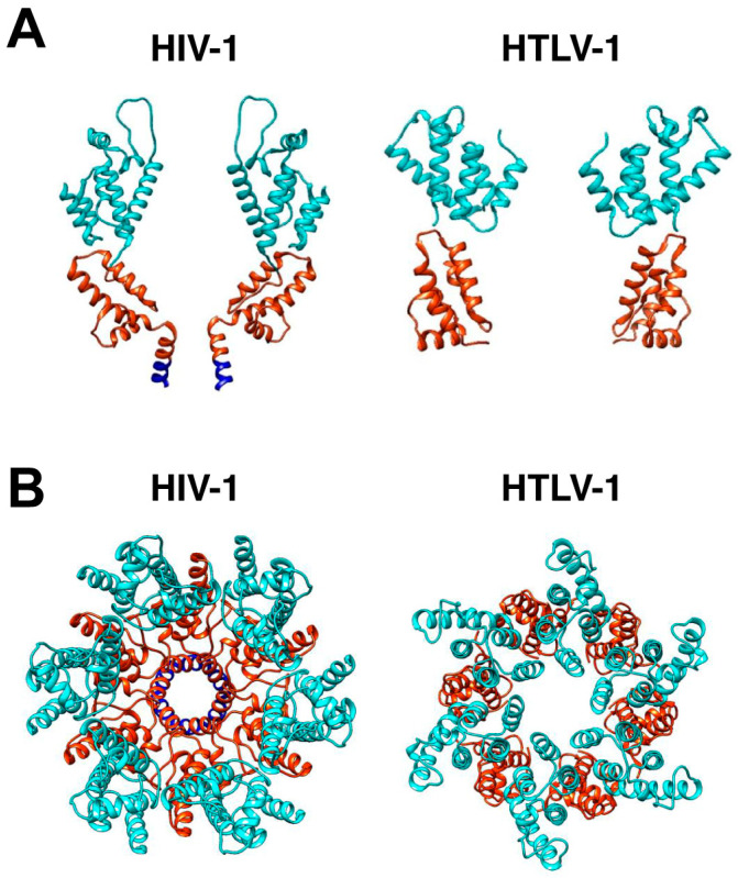
Figure 4.
HIV-1 and HTLV-1 Gag hexamer structures. (A) The two HIV-1 CA molecules are displayed on the side of the hexamer, with CANTD in cyan and CACTD in orange. HIV-1 SP1 domains are shown in blue. The PDB codes are HIV-1 (5L93) [109], HTLV-1 CANTD (8PUG) [183], and HTLV-1 CACTD (8PUH) [183]. The cross-section of the HTLV-1 Gag lattice reconstruction map suggests a distinctive arrangement of the CANTD and CACTD compared to HIV-1. (B) Shown is the top view of the HIV-1 hexamer structure, which was generated by fitting HIV-1 CA (5L93) into the EM density of the immature HIV-1 lattice (EMD: 4017). The top view of the HTLV-1 Gag hexamer structure shown was generated by fitting CANTD and CACTD separately into the EM density of the immature HTLV-1 CA lattice (EMD: 17942). The flexible linker between HTLV-1 CANTD and CACTD is unstructured and is therefore not shown.

