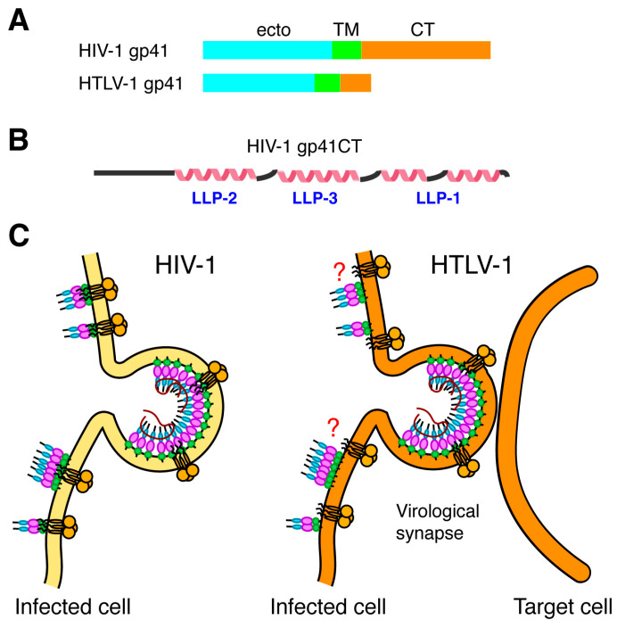
Figure 5.
Comparison of Env CT. (A) Schematic representation of the gp41 subunits, indicating the lengths of their respective cytoplasmic tails (25 and 150 amino acids for HTLV-1 and HIV-1, respectively. (B) Secondary structure representation of the HIV-1 gp41CT protein based on the NMR data [192]. (C) HIV-1 Env incorporation is mediated by interaction between the MA domain of the Gag lattice and gp41CT. For HTLV-1, the CT appears to contain functional motifs that play important roles in cell-to-cell infection and syncytium formation.

