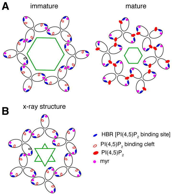
Figure 6.
Comparison of MA lattices based on structural data. (A) Schematic representation of the myrMA lattice in the immature and mature states based on the cryo-ET data [170]. The trimer–trimer interactions are mediated by the N-terminal domain in the vicinity of the myr group, while the PI(4,5)P2 binding pocket is empty. In the mature myrMA lattice, PI(4,5)P2 is bound to the cleft and myrMA trimer–trimer interactions are formed by the HBR and PI(4,5)P2. (B) Schematic illustration of the myrMA lattice based on the X-ray structure of myrMA. In this lattice, myrMA–myrMA interaction at the trimer–trimer interface is mediated by the N-terminal residues. Of note, myrMA–myrMA interaction at the trimer–trimer interface places the myr groups (red) in juxtaposition. The HBR and PI(4,5)P2 binding cleft are also shown. Hexagons and triangles denote C6 and C3 symmetry, respectively.

