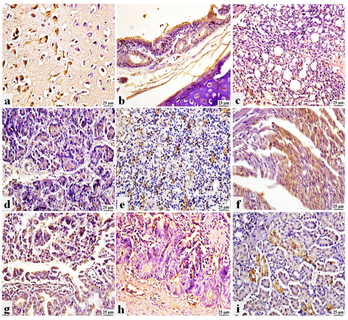
Figure 6.
Photomicrograph of immunohistochemistry (IHC) of PPMV-1 presence in different tissue organs of non-vaccinated/challenged pigeons. (a) Brain: positive expression in degenerated neurons. (b) Trachea: positive expression in the tracheal epithelium and in the inflammatory cells of the propria submucosa. (c) Lung: positive expression in air capillaries. (d) Pancreas: positive expression in the exocrine epithelial cells. (e) Spleen: positive expression in the splenic parenchyma. (f) Heart: positive expression in the cardiac muscle. (g) Proventriculus: positive expression in the glandular epithelium and desquamated epithelial cells. (h) Intestine: positive expression in intestinal mucosa. (i) Kidneys: positive expression in the renal tubular epithelium.

