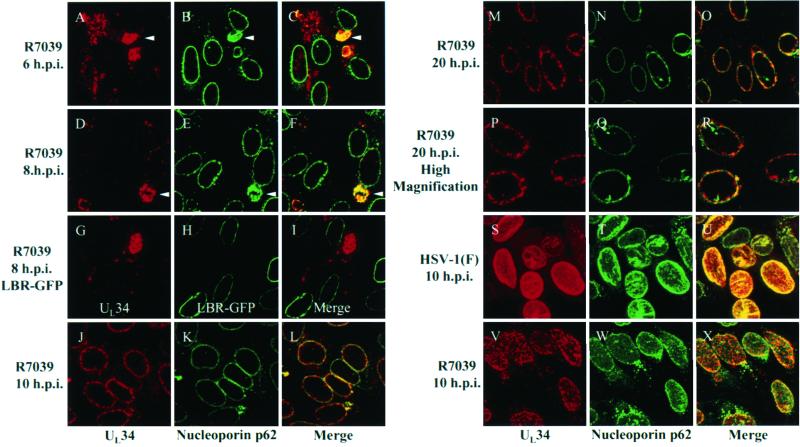
FIG. 7.
Digital confocal images showing localization of UL34 protein in cells infected with a US3 null virus. (A to F and J to R) HEp-2 cells were infected at an MOI of 10 with US3 null HSV-1(F) (R7039) for 6 h (A to C), 8 h (D to F), 10 h (J to L), and 20 h (M to R). Infected cells were fixed with formaldehyde and immunostained with chicken anti-UL34 antibody detected with donkey anti-chicken Ig-Texas Red conjugate and with mouse anti-nucleoporin p62 detected with goat anti-mouse Ig-FITC conjugate. (G to I) HEp-2 cells were transfected with an LBR-GFP expression construct and allowed to express for 24 h. Transfected cells were subsequently infected for 8 h with US3 null virus R7039, fixed with formaldehyde, and immunostained with chicken anti-UL34 detected with donkey anti-chicken Ig-Texas Red conjugate. (S to X) HEp-2 cells were infected with HSV-1(F) (S to U) or R7039 (V to X) at an MOI of 10 for 10 h fixed, permeabilized, and immunostained with anti-UL34 and anti-nucleoporin p62. Confocal Z-sections are shown in panels A to R, and confocal Z-stacks are shown in panels S to X. Arrows in panels A through F indicate positions of perinuclear UL34-nucleoporin p62 structures. Original magnifications: A to O and S to X, X630; P to R, X1,000.

