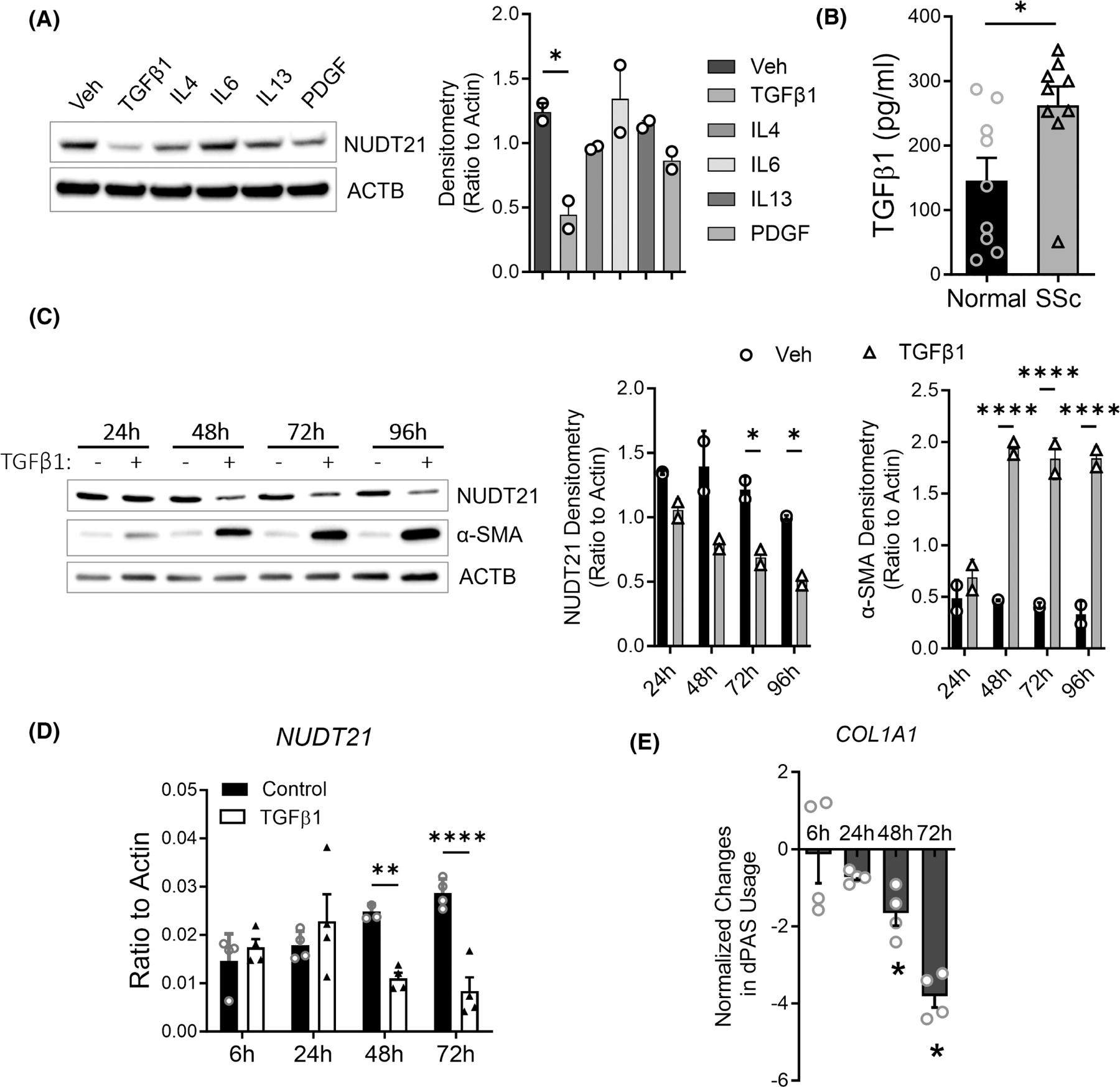
FIGURE 1.

Transforming growth factor beta 1 (TGFβ1) downregulates NUDT21 in dermal fibroblasts. (A) Left panel: Western blot of NUDT21 and β-Actin (ACTB) was performed using the protein lysate of normal human dermal fibroblasts treated with PBS, 5 ng/mL TGFβ1, 10 ng/mL interleukin 4 (IL4), 60 ng/mL IL6 + interleukin 6 receptor (IL6R), 10 ng/mL IL13, or 30 μL/mL platelet-derived growth factor (PDGF) for 2 days. Right panel: Densitometry analysis of the Western blot image. (B) The levels of TGFβ1 in the supernatant of cultured normal or SSc dermal fibroblasts were determined using ELISA. (C) Left panel: The protein levels of NUDT21 were determined in normal human dermal fibroblasts treated with 5 ng/mL TGFβ1 for different hours. Right panels: Densitometry analysis of the Western blot image. (D) The transcript levels of NUDT21 in normal human dermal fibroblasts treated with 5 ng/mL TGFβ1 for different hours. (E) The normalized percentage of distal polyadenylation site (dPAS) usage for COL1A1 was determined using PCR methods, and data were presented as Log (% long transcript in TGFβ1 cells/% long transcript in control) ± mean square error (MSE). p-values were calculated using a two-tailed student t-test for panel B, ANOVA followed by Dunnett’s multiple comparisons test for panels A, C, and D, and one sample t-test versus 0 for panel C. *p < .05; **p < .01; ****p < .0001.
