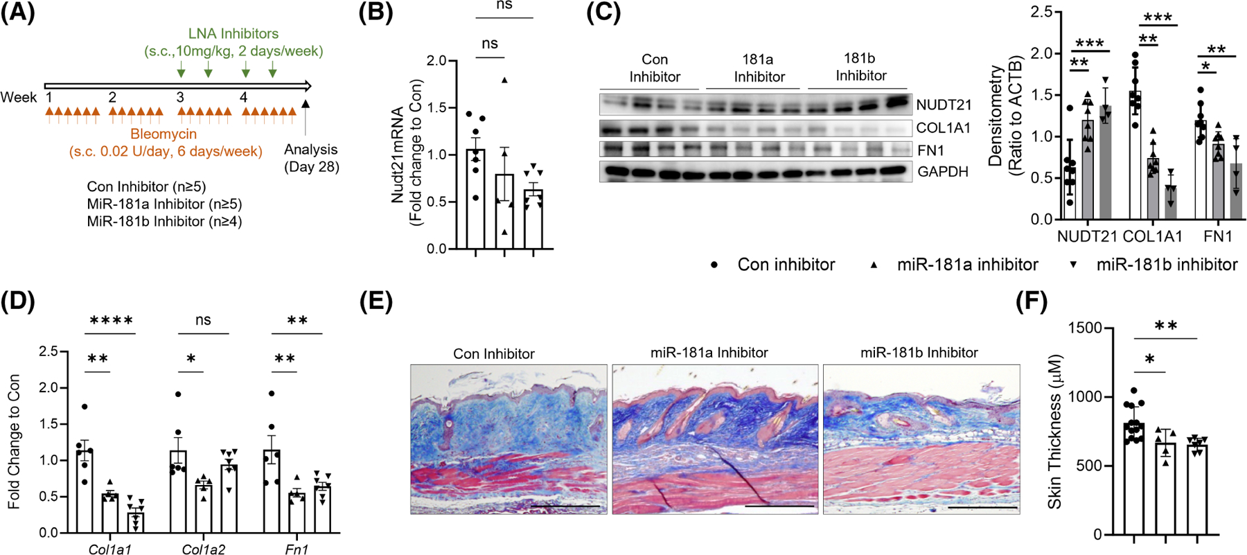
FIGURE 5.

Protective roles of miR-181a and miR-181b inhibitors in skin fibrosis. (A) Scheme of experimental design. Eight-week-old female wild-type C57/BL6 mice were injected with repeated s.c. bleomycin six times a week for 4 weeks. Two weeks after the first bleomycin injection, mice were s.c. injected with 10 mg/kg/day LNA control, miR-181a, or miR-181b inhibitors twice a week at the bleomycin-injected spot. The skin was collected 28 days after the first bleomycin injection for analysis. (B, D) Real-time qRT PCR was performed to determine the transcript levels of (B) Nudt21, and (D) Col1a1, Col1a2, and Fn1. (C) Left panel: Western blot was used to determine the protein levels of NUDT21, COLIA1, FN1, and ACTB. Right panel: Densitometry analysis of the Western blot image. n ≥ 4 fibroblast lines from different mice. (E) Masson’s Trichrome staining was performed to show dermal fibrosis in different treatment groups. Scale bar = 50 μm. (F) Quantification of dermal thickness. N ≥ 5. The graphs represent means ± MSE. p-value was determined using one-way ANOVA (C, F) or two-way ANOVA (D) followed by Dunnett’s multiple comparisons tests. *p < .05; **p < .01; ***p < .001; and ****p < .0001.
