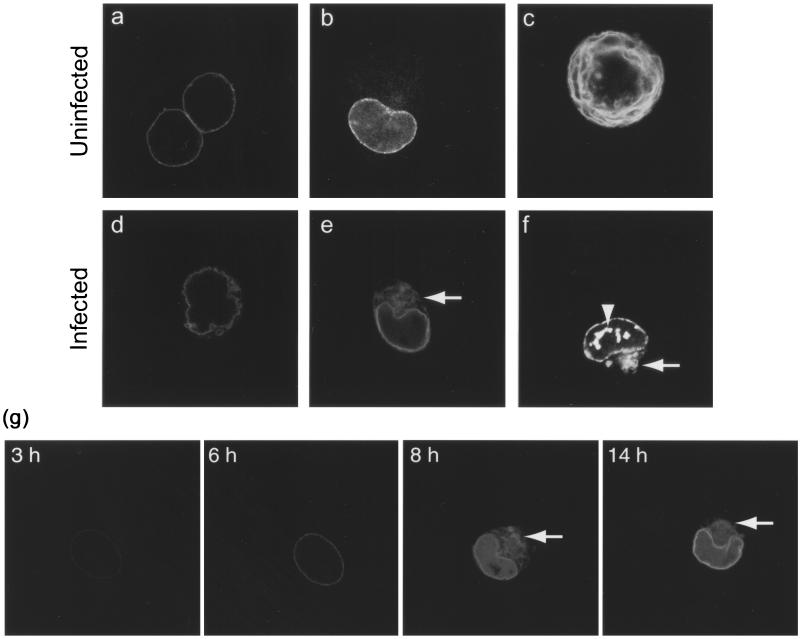
FIG. 1.
Live COS-1 cells transiently expressing LBR-GFP viewed by confocal microscopy. (a and b) Uninfected interphase cells show nuclear-rim fluorescence and some fluorescence in the ER (b). (c) Uninfected mitotic cell shows fluorescence in the ER. (d to g) Cells infected with HSV-1 at 10 PFU/cell (e, 8 h; d and f, 24 h) show a distorted nuclear-rim pattern (d) in a membranous cytoplasmic compartment (e and f [arrows]) and in intranuclear domains (f [arrowhead]). (g) A single COS-1 cell transiently expressing LBR-GFP and infected with HSV-1 at 10 PFU/cell was tracked throughout the course of infection. Live confocal microscope images are shown, taken between 3 and 14 h p.i. Between 6 and 8 h p.i., the nuclear-rim LBR-GFP partially redistributes to a cytoplasmic compartment (arrows) which is stable over several hours.

