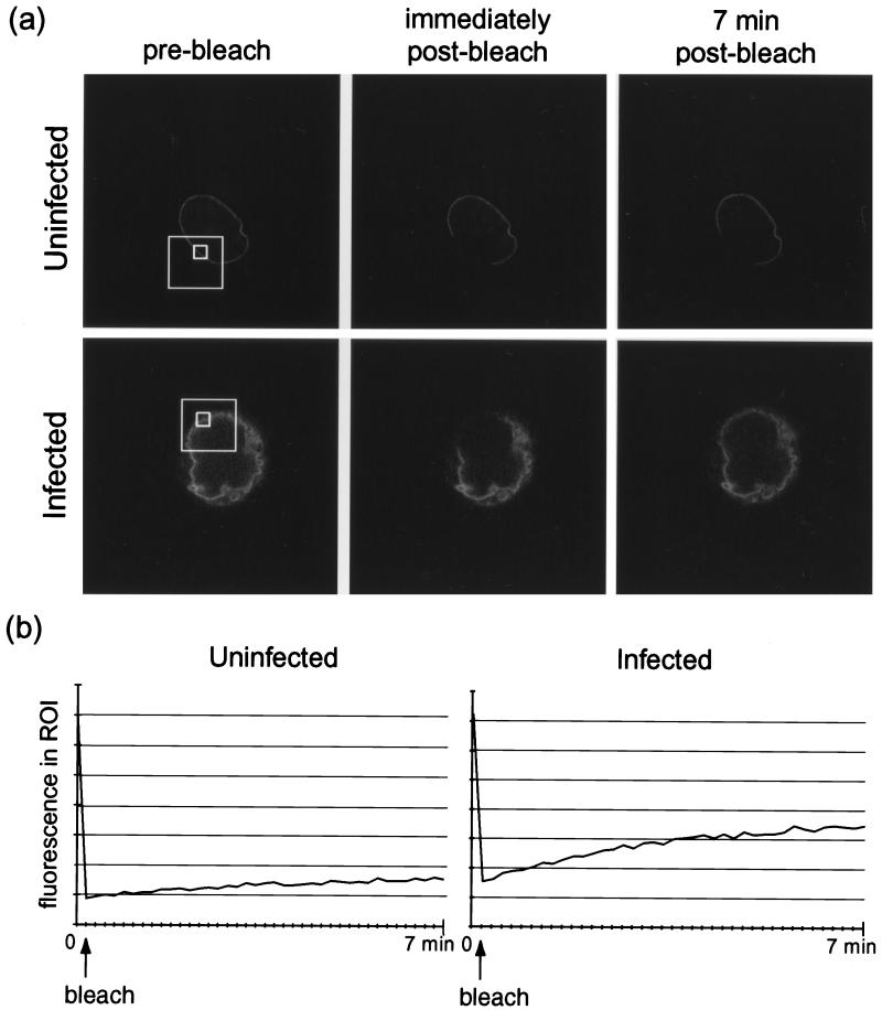
FIG. 4.
COS-1 cells transiently expressing LBR-GFP were infected with HSV-1 at 10 PFU/cell or left uninfected and viewed live on a confocal microscope at 24 h p.i. (a) A restricted region (large frames) of the nuclear-rim fluorescence was irreversibly photobleached by a 10-s, 100% intensity laser pulse. Images of the whole cell were recorded before bleaching and at 10-s intervals for 7 min following the bleaching; selected images are shown. (b) LSM software was used to record fluorescence (in arbitrary units) in ROIs within the bleached area (panel a, small frames). These readings from the cells shown in panel a are plotted over time.

