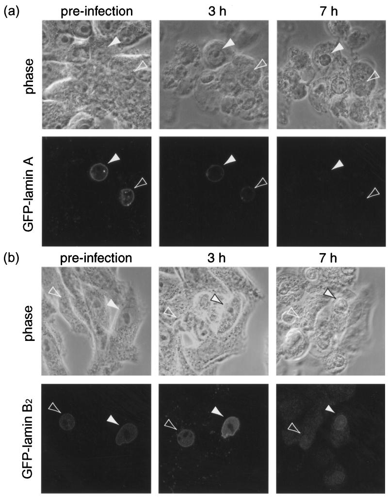
FIG. 8.
Examples of individual BHK-21 cells stably expressing GFP-lamin A (a) or GFP-lamin B2 (b) and infected with HSV-1 at 10 PFU/cell were followed by phase and fluorescence confocal microscopy during the course of infection. The arrowheads indicate cells expressing GFP-lamin which exhibit CPE and reduced nuclear-rim fluorescence as the infection progresses.

