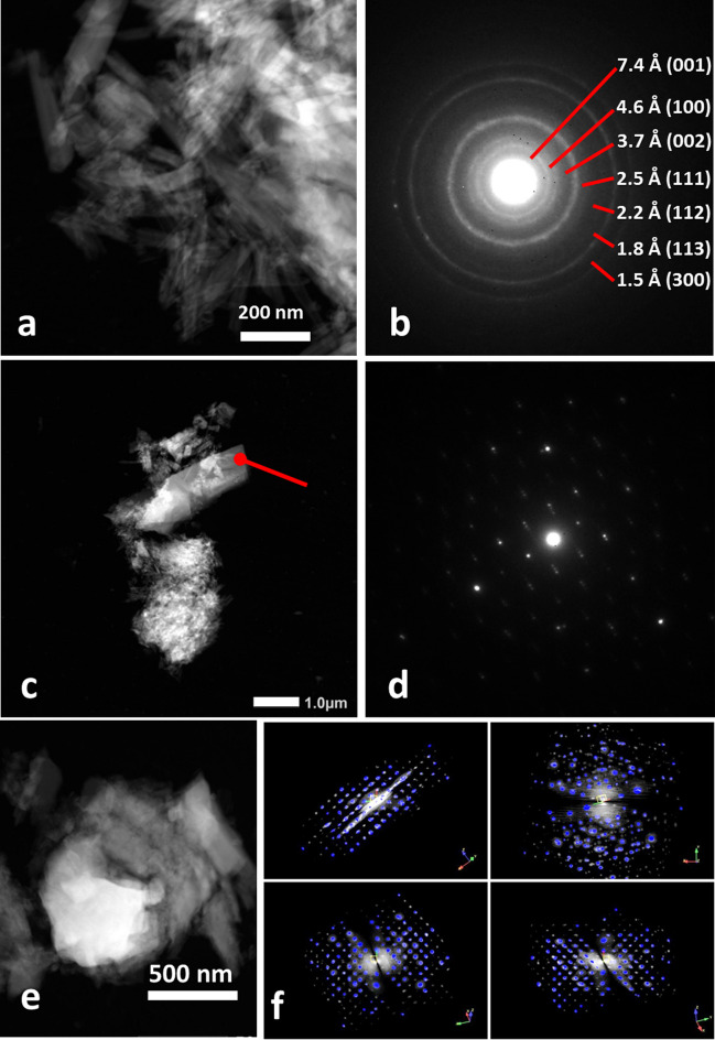
Fig. 4.
TEM analysis of NSFe showing a composition of serpentine polymorphs and magnetite: a chrysotile fibers with 13.5% Fe; b electron diffraction pattern of polycrystalline area in a; c modulated antigorite with 3.84% Fe; d selected area electron diffraction (SAED) of the spot highlighted in c; e magnetite, Fe3O4 (76% Fe, close to stoichiometric 72%); f 3D electron diffraction of the cubic face-centered cell of magnetite, with a = 8.4 Å

