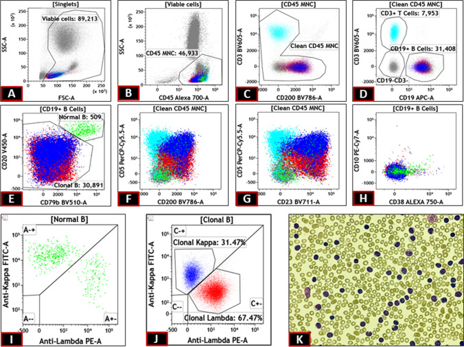
Fig. 1.
Mononuclear cell were gated on SSC Vs CD45 plot (B) after removal of debris on SSC Vs FSC (A). CD19 positive cells were gated (D) after removal of junk on CD3 Vs CD200 plot(C). Normal B cells were identified as bright CD20 and CD79b positive cells (E). The rest abnormal B cells showed immunophenotype consistent with CLL (E to H) but with split on kappa lambda plot (J) but with dimmer intensity than the normal B cells (I). Peripheral smear showing small cell lymphocytosis (K)

