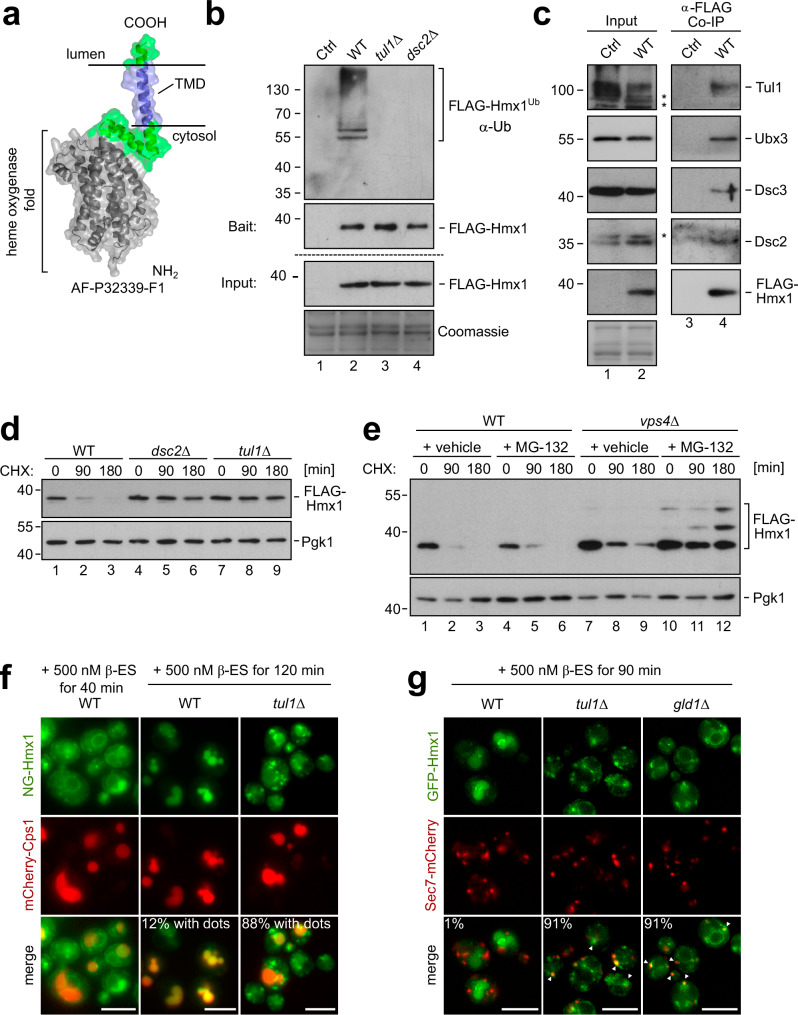
Fig. 3. The heme oxygenase 1 (Hmx1) is ubiquitinated by the Dsc complex and degraded by ESCRT – and EGAD pathways.
a AF model of Hmx1 (AF-P32339-F1). b–e SDS–PAGE and Western blot analysis with the indicated antibodies: b, c Input and elution of b denaturing or c non-denaturing FLAG-Hmx1 immunoprecipitations (IP) from indicated cells. Control (Ctrl) cells were untagged WT strains. Representative blots from 3 independent experiments with similar results are shown ‘*’ unspecific antibody cross-reactions; d, e Total cell lysates of the indicated cells that were untreated (0 min) or treated with 50 µg/mL cycloheximide (CHX) to block protein synthesis for the indicated times. Pgk1 served as a loading control. Densitometric quantification is shown in Supplementary Fig. 3a. f, g Live cell epifluorescence microscopy of the indicated cells, induced with 500 nM ß-estradiol for the indicated times. f mNeongreen-ALFA-Hmx1 (NG-Hmx1) (green) and mCherry-Cps1 (red). Indicated are % of WT cells (n = 110) or tul1Δ cells (n = 119) in which dots outside of vacuoles was detected. g Co-localization of induced GFP-Hmx1 (green) with Sec7-mCherry (red). Indicated are % of WT cells (n = 80), tul1Δ cells (n = 82), gld1Δ cells (n = 58) in which co-localization of GFP-Hmx1 and Sec7 was detected. Scale bars 5 µm. See also Supplementary Fig. 3.

