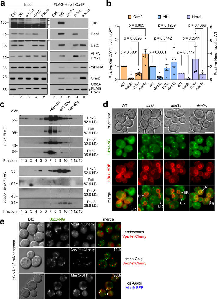
Fig. 5. Dsc2 is required for substrate recognition.
a, c SDS–PAGE and Western blot analysis with the indicated antibodies: a elution and input from non-denaturing Ubx3-Flag immunoprecipitations from the indicated cells. Control (Ctrl) cells were untagged WT strains. Representative blots from 3 independent experiments with similar results are shown. b Densitometric quantification of the indicated co-immunoprecipitated substrate proteins Orm2, Yif1: WT, dsc2Δ (n = 7 independent experiments), tul1Δ, and dsc3Δ (n = 5 independent experiments); Hmx1 (n =n = 3 independent experiments). Data are presented as mean ± standard error of the mean (SEM). Data were analyzed by a two-tailed paired t-test. c Ubx3-Flag was immunoprecipitated from WT- or dsc3Δ cells and immunoprecipitated proteins were subjected to size exclusion chromatography. Mr (in kDa) of the proteins and the standards are indicated. d, e Live cell epifluorescence of WT and the indicated mutants expressing Ubx3-mNG (green) with d dsRED-HDEL (red). ER indicated. e Vps4-mCherry, Sec7-mCherry or Mnn9-BFP. In 93% of cells (n = 75) Ubx3-NG partially co-localized with or localized in the vicinity of Mnn9-BFP and in 14% of cells (n = 58) Ubx3-NG partially co-localized with or localized in the vicinity of Sec7-mCherry. Scale bars 5 µm.

