Figure 3.
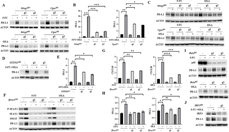
5-FU/oxaliplatin upregulates PD-L1 expression in a cGAS/STING/IFNβ-dependent manner in colon cancer cells. (A, B) Sting1 or Cgas knockout (Sting1KO or CgasKO ) MC38 cells were treated with 2 μM 5-FU or 20 μM oxaliplatin (OXA) (A) or 2 μM 5-FU plus 10 μM oxaliplatin (OXA) (B) for 24 hours. Western blot (A) and Q-PCR (B) analysis were performed to examine PD-L1 expression. (C) Sting1 or Cgas knockout (Sting1KO or CgasKO ) CT26 cells were treated with 2 μM 5-FU or 20 μM oxaliplatin (OXA) for 24 hours. Western blot analysis was performed to examine PD-L1 expression. (D, E), STING1 knockout (STING1KO ) HT29 cells were treated with 5 μM 5-FU plus 20 μM oxaliplatin (OXA) for 48 hours. Western blot (D) and Q-PCR (E) analysis were performed to examine PD-L1 expression. (F-H) Ifnar1 knockout (Ifnar1KO ) MC38 cells were treated with 2 μM 5-FU or 20 μM oxaliplatin (OXA) for 24 hours. Western blot analysis was performed to examine expression of STAT1, P-STAT1 (Y701), ISG15 and PD-L1 expression (F). Expression of CXCL10 (G) and PD-L1 (H) was determined by Q-PCR analysis. (I) Rela knockout (RelaKO ) MC38 cells were treated with 2 μM 5-FU (upper panel) or 20 μM oxaliplatin (OXA, lower panel) for 24 hours. Western blot analysis was performed to examine expression of p65 and PD-L1. (J) IRF3 knockout (IRF3KO ) HT29 cells were treated with 5 μM 5-FU plus 20 μM oxaliplatin (OXA) for 48 hours. Western blot analysis was performed to examine expression of IRF3 and PD-L1. *P<0.05, **P<0.01, ***P<0.001, ****P<0.0001.

