Figure 5.
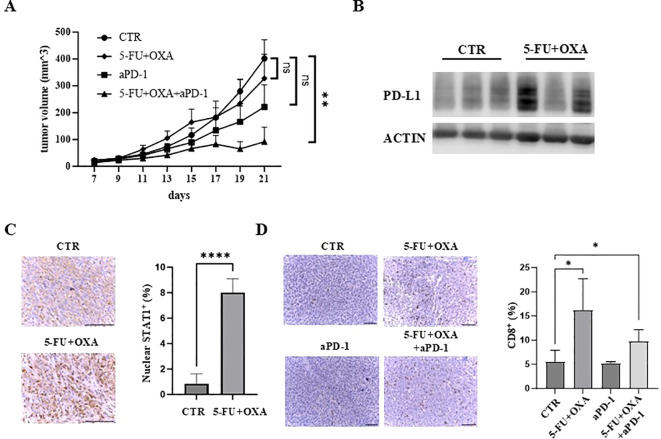
The combination of 5-FU/oxaliplatin and anti-PD-1 treatment efficiently inhibit tumor growth in vivo. (A) Tumor growth curves of MC38 cells treated with saline/a control IgG (CTR), 5-FU (25 mg/kg) plus oxaliplatin (OXA, 2.5 mg/kg) (5-FU+OXA), an anti-PD-1 antibody (200 μg) (aPD-1) or 5-FU (25 mg/kg)/oxaliplatin (OXA, 2.5 mg/kg) plus an anti-PD-1 antibody (200 μg) (5-FU+OXA+aPD-1) are shown. N = 5-10. (B) Western blot analysis was performed to examine PD-L1 expression in vehicle- (CTR) or 5-FU/oxaliplatin-treated (5-FU+OXA) tumors. (C) Representative images of IHC staining of STAT1 in control (CTR) and 5-FU/oxaliplatin (5-FU+OXA) treated tumors. Scale bars: 100 μm (left). Quantification of percentage of nuclear STAT1-positive cells is shown in the right panel. (D) Representative images of IHC staining of CD8 in tumors of each group. Scale bars: 100 μm (left). Quantification of percentage of CD8-positive cells is shown in the right panel. *P<0.05, **P<0.01, ****P<0.0001.

