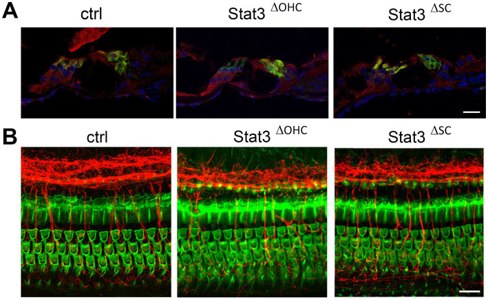
Figure 1.
Immunohistochemical analyzation of Stat3ΔOHC and Stat3ΔSC. (A) Stat3 (red) is highly expressed in sensory hair cells (green, Myo7a) or supporting cells in control cochleae, but is reduced in OHC in Stat3ΔOHC and in supporting cells of the Stat3ΔSC mice cochleae. (B) However, immunohistochemical analyzation of the cochlear duct indicated no cell loss of OHC (green, Phalloidin), altered innervation (red, βIII-tubulin) or morphology of the sensory epithelium of the cochlea. Ctrl: n = 3, Stat3ΔSC: n = 3. Scale bars: 20 μm.

