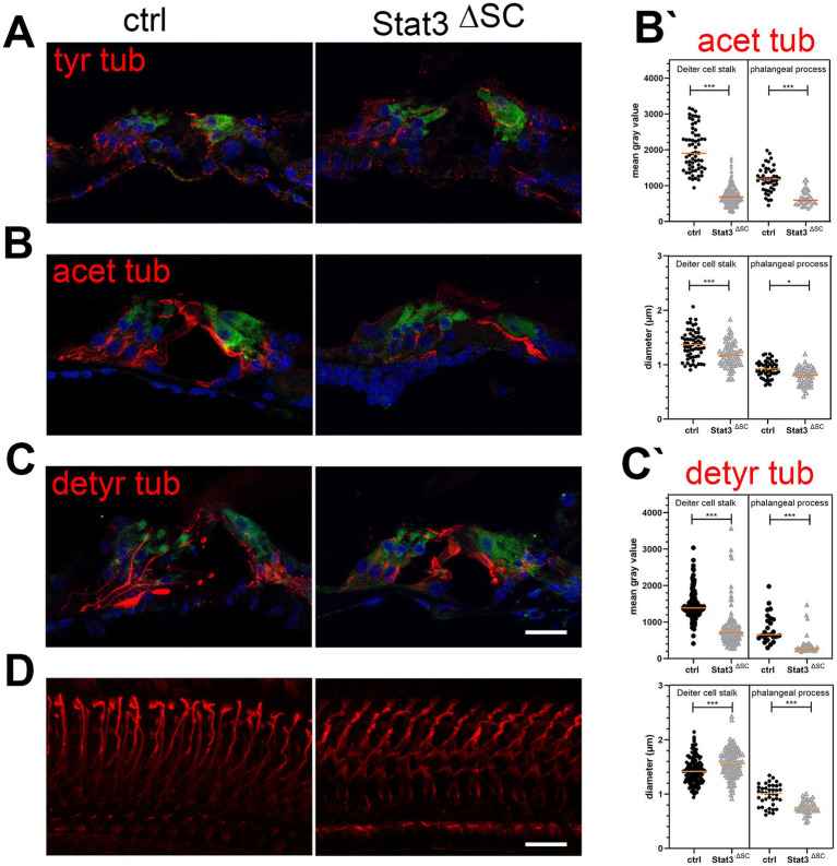
Figure 4.
Immunohistological analysis of post-translational modifications of microtubules in Stat3ΔSC organ of Corti cryosections with OHC marker Myosin 7a (green). (A) Tyrosinated tubulin (tyr tub, red) is expressed on sensory hair cells, nerve fibers and supporting cells of the sensory epithelium of the cochlea. (B) Acetylated tubulin (acetyl tub, red) is expressed in Deiters and pillar cells, but are significantly reduced in Stat3ΔSC. (B′) Statistical analyzation of acetylated tubulin in mean gray value and the diameter of Deiters cells stalk base and phalangeal processes showed decreased acetylated tubulin (p < 0.001) and reduced diameter in the stalk base (p < 0.001) and phalangeal processes (p < 0.05) inStat3ΔSC. (C) Detyrosinated tubulin (detyr tub, red) is expressed in Deiters and pillar cells, but are significantly reduced in Stat3ΔSC. (C′) Statistical analyzation of detyrosinated tubulin in mean gray value of Deiters cells stalk base and phalangeal processes showed decreased acetylated tubulin (p < 0.001). However, diameter of Deiters cells stalk of Stat3ΔSC is significantly higher compared to control Deiters cells (p < 0.001), whereas the phalangeal processes of Stat3ΔSC is significantly reduced compared to control phalangeal processes. (D) Whole Mount staining with detyrosinated tubulin revealed well-defined structures of detyrosinated microtubule bundles in control cochlea, whereas these modified bundles show progressive instability with high tortuosity. Statistical analyses: Unpaired t-test, two-tailed. Ctrl: n = 3, Stat3ΔSC: n = 3. Scale bars: 20 μm. Significances: *p < 0.05, ***p < 0.001.

