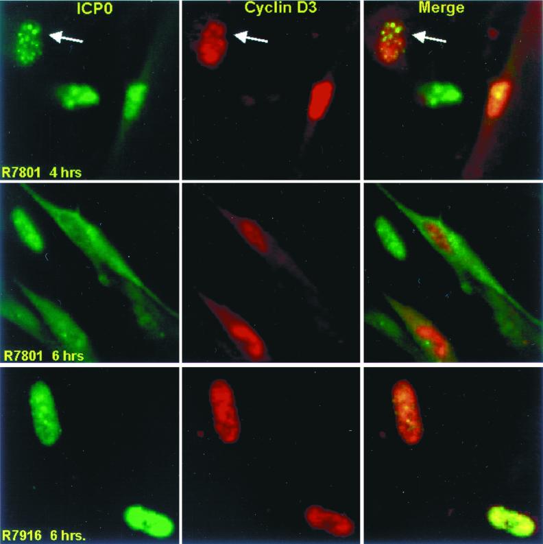
FIG. 11.
Digital images of HEL fibroblasts infected with wild-type (R7801) and D199A mutant (R7916) viruses expressing cyclin D3 and reacted with antibodies to ICP0 and cyclin D3. The infected cells were fixed 4 or 6 h after infection and reacted with antibodies and processed as described in the legend to Fig. 10. The digital images were not modified subsequent to capture. The arrows point to ICP0 and cyclin D3 colocalized in the nucleus of the cell shown in the upper left corner of each panel.

