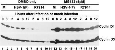
FIG. 4.
Photographs of immunoblots of electrophoretically separated quiescent HEL cell lysates treated with proteasomal inhibitor MG132 (lanes 11 to 20) or only with DMSO (lanes 1 to 10). Quiescent HEL cells were either mock infected (lanes 1 and 2 and 11 and 12) or infected with HSV-1(F) (lanes 3 to 6 and 13 to 16) or R7914 (lanes 7 to 10 and 17 to 20). The lysates were harvested at the times indicated, subjected to electrophoresis on SDS-12% polyacrylamide gels, transferred to nitrocellulose, and reacted with mouse monoclonal antibodies against cyclins D1 and D3.

