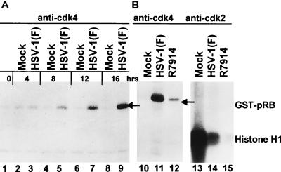
FIG. 5.
(A) Autoradiographic image of electrophoretically separated [γ-32P]ATP-labeled GST-pRB fusion protein. Synchronized HeLa cells were mock infected (lanes 1, 2, 4, 6, and 8) or infected with HSV-1(F) (lanes 3, 5, 7, and 9). The cells were harvested at the times indicated, solubilized, electrophoretically separated in denaturing gel, transferred to a nitrocellulose sheet, and reacted with polyclonal anti-cdk4. Immunoprecipitates were incubated with the substrate GST-pRB in a kinase buffer supplemented with [γ-32P]ATP. The reaction mixtures were subjected to electrophoresis in a denaturing 10% polyacrylamide gel, transferred to a nitrocellulose membrane, and subjected to autoradiography. The radiolabeled product was collected on a phosphorimager. (B) Lanes 10 to 12, autoradiographic image of electrophoretically separated GST-pRB labeled with [γ-32P]ATP by cdk4 immune precipitated from synchronized HeLa cells harvested 12 h after mock infection (lane 10) or infection with HSV-1(F) (lane 11) or R7914 (lane 12). Experimental details were the same as for panel A. Lanes 13 to 15, autoradiographic image of electrophoretically separated histone H1 labeled with [γ-32P]ATP by cdk2 immune precipitated from synchronized HeLa cells 12 h after mock infection (lane 13) or infection with HSV-1(F) (lane 14) or with R7914 (lane 15). Experimental details were the same as for panel A.

