Fig. 6.

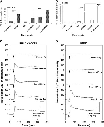
Mast cell stimulation is highest upon co-stimulation of FcεRI and CCR1 as assessed by functional assays. Percent β-hexosaminidase release was increased in (A) RBL-CCR1 cells and (B) BMMC following DNP-HSA cross-linking of FcεRI and/or stimulation of CCR1 with MIP-1α for 30 min at 37°C. Degranulation was greatest when cells were co-stimulated with both ligands. Assays were performed twice independently, with conditions tested in triplicate using 40 000 BMMC (6 weeks old) per well. Data from one typical experiment are shown, ***P < 0.0001. To assess calcium mobilization, intact RBL-CCR1 cells (C) and 7-week BMMC (D) were sensitized with 10 ng ml−1 or 100 ng ml−1 anti-DNP IgE (SPE-7), loaded with Indo 1-AM, and stimulated with antigen (10 ng ml−1 DNP-HSA) and/or MIP-1α (50 ng ml−1). The intracellular rise in calcium was immediately observed from 0 s to 5 min. Traces are representative of two independent experiments.
