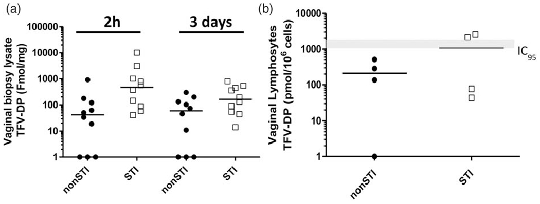
Fig. 4. Tissue TFV-DP concentrations and median (bar) following vaginal dosing with 1% TFV gel.

STI, sexually transmitted infection; TFV-DP, tenofovir diphosphate. (a) Vaginal tissue homogenates 2 h and 3 days after vaginal TFV gel dosing. (b) Vaginal lymphocytes from STI-negative (closed circles) and STI-infected (open squares) animals 3 days after vaginal TFV dosing. Solid black line indicates median TFV-DP concentrations. Grey line indicates TFV-DP concentrations that correspond to the in-vitro 95% inhibitory concentration (IC95) range [9].
