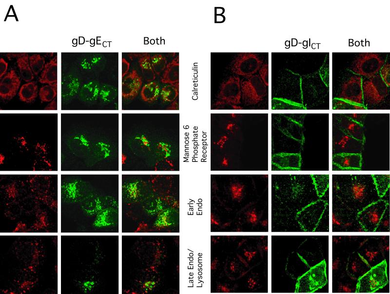
FIG. 4.
Subcellular localization of gD-gECT and gD-gICT compared with that of other cellular markers. HEC-1A cells were coinfected with either AdgD-gECT and Ad-Trans (A) or AdgD-gICT and Ad-Trans (B) as described in the legend to Fig. 3. The cells were stained with anti-gD MAb DL6 (green) and rabbit anticalreticulin antibodies or rabbit anti-MPR antibody (red) followed by Alexa 594-conjugated goat anti-rabbit IgG and Alexa 488-conjugated goat anti mouse IgG. Staining for early endosomes involved incubation with Alexa 594-conjugated Tf (red) for 15 min at 37°C, followed by washing and immediate fixation. Late endosomes/lysosomes were stained as for early endosomes, except that there was a further incubation at 37°C for 30 min.

