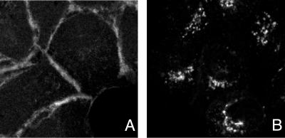
FIG. 5.
gE/gI accumulate in the TGN, rather than at cell junctions, in Ad-infected cells. HEC-1A cells were infected with Ad(E1−)gE and Ad(E1−)gI for 24 h and then stained by one of two methods. In panel A, the cells were stained essentially as previously described (17). The cells were incubated with a mouse MAb specific for β-catenin, followed by Alexa 594-conjugated goat anti-mouse IgG antibodies. The cells were washed and incubated with Oregon Green (a fluorescein analog) conjugated anti-gE MAb 3114, washed, and incubated with goat anti-fluorescein antibodies conjugated to Alexa 488. In panel B, the same protocol was followed except that following the Alexa 594-conjugated goat anti-mouse IgG antibodies, the cells were incubated with 2% mouse serum for 2 h, in order to prevent the anti-mouse antibodies from reacting with MAb 3114.

