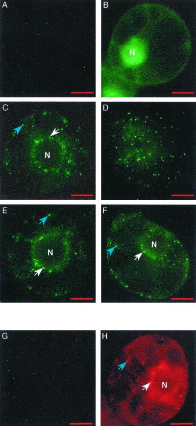
FIG. 7.
Localization of P15 in tobacco BY-2 protoplasts. Protoplasts were mock inoculated (A and G) or inoculated with T1 + TRep-EG (B), T1-EG15 + T2 (C and D), T1 + TRep-EG15 (E), pCK-EG15 (F), or viral RNA (H). Observations were carried out 48 hpi. EGFP expression (A to F) and immunostaining of P15, using P15 antiserum and Cy3 antibodies (G and H), were detected by epifluorescence microscopy. White arrows indicate a localization of P15 around the nucleus (N), and blue arrows indicate a localization within the cytoplasm. 4′,6′-Diamidino-2-phenylindole (DAPI) staining (not shown) was used to localize the nucleus. For panel F, the number of integrated images was increased to obtain fluorescence comparable to that of panels C to E. Bar, 10 μm.

