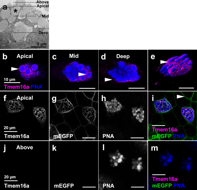
Fig. 3.
Tmem16a localises to the plasma and not vesicle membrane in SSCs. (a) SEM of SSCs reveals the presence of large vesicles (example indicated by asterisk) that bulge beyond the apical cell membrane of SSCs. Dashed lines indicate the planes of view in (b–m). (b–e) Immunofluorescent localisation of Tmem16a protein in apical, mid and basal planes of a single SSC reveals Tmem16a is present in a pattern that, in the apical (b) and mid (c) planes, appears to surround the PNA-positive secretory vesicles regions of cell (arrowheads). However, this localisation pattern is absent around vesicles in the more basal region of the cell (d), and expression becomes evident in the presumed plasma membrane (d, arrowhead). 3D surface rendering shows Tmem16a at an apical plane below that of the bulging vesicles (e). (f-i) In the apical plane of epidermal cells, Tmem16a (f) localises with mEGFP (g) in SSCs stained with PNA (h; merged in i), but does not localise with mEGFP marking the plasma membrane of other cell types (h, arrowhead). (j–m) Above the plane of epidermal cells, Tmem16a (j) and mEGFP (k) immunofluorescence is absent from bulging PNA-positive secretory vesicles (l; merged in m), demonstrating that Tmem16a is absent from the secretory vesicle membrane.

