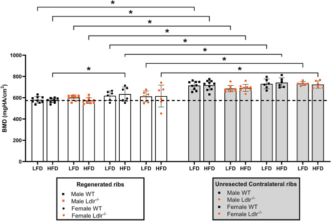
Fig. 4.
BMD remains unchanged between male and female mice under HFD. Bone mineral density (BMD—mg HA/cm3) of (A) regenerated rib callus and (B) its respective unresected contralateral rib in each mouse group at 21 dpr. The dashed line represents the overall mean value of the group of male wild-type (WT) mice under low-fat diet (LFD) and was used as a reference for comparison with all other groups. All BMD measurements were obtained from ex vivo high resolution micro-computed tomography data and analyzed using AnalyzePro 14.0 software. Data are displayed as the mean ± SD, considering WT male mice under LFD (n = 9), WT male mice under high-fat diet (HFD) (n = 10), Ldlr−/− male mice under LFD (n = 9), Ldlr−/− male mice under HFD (n = 9), WT female mice under LFD (n = 6), WT female mice under HFD (n = 6), Ldlr−/− female mice under LFD (n = 6), and Ldlr−/− female mice under HFD (n = 6). Statistical analysis was performed using two-way analysis of variance (ANOVA) followed by Fisher’s LSD test. Significant differences between groups are expressed as p < 0.05. dpr = days post-resection; high-fat diet (HFD).

