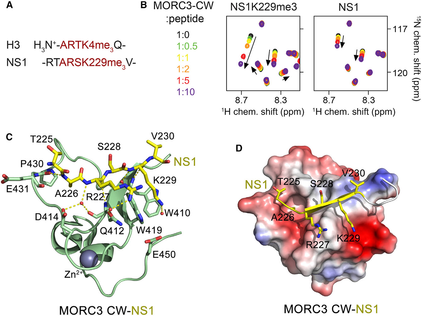
Figure 1. MORC3-CW Is a Target of NS1.

(A) The amino-terminal sequence of H3 (trimethylated at lysine 4) and the C-terminal sequence of NS1 (trimethylated at lysine 229) are shown.
(B) Superimposed 1H,15N HSQC spectra of 15N-labeled MORC3-CW collected upon titration with the methylated and unmodified NS1 peptides. Spectra are color coded according to the protein-to-peptide molar ratio.
(C) The crystal structure of the MORC3-CW:NS1 complex. The CW domain is shown in a ribbon diagram (green), and NS1 is shown as sticks (yellow). The zinc (gray) atom is shown as a sphere.
(D) Electrostatic surface potential of the MORC3-CW domain colored blue and red for positive and negative charges, respectively. The bound NS1 peptide is yellow.
See also Figure S1.
