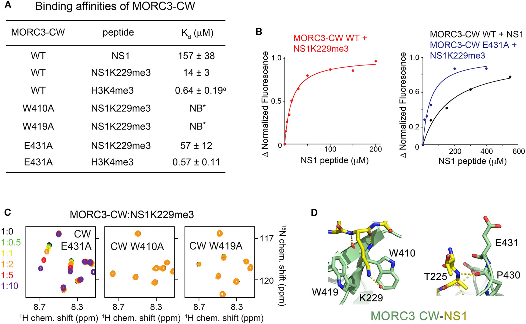
Figure 2. Mechanistic Insight into H3 Mimicry by NS1.

(A) Binding affinities of WT and mutated MORC3-CW to indicated peptides as measured by tryptophan fluorescence or NMR (*). The binding affinity of the CW domain to H3K4me3 peptide was determined previously (a) (Andrews et al., 2016). The experiments were carried out in triplicate (in duplicate for the CW E431A-H3K4me3 interaction). Error bars denote SD.
(B) Representative binding curves used to determine the Kd values by tryptophan fluorescence in (A).
(C) Superimposed 1H,15N HSQC spectra of the 15N-labeled MORC3-CW mutants collected upon titration with the NS1K229me3 peptide. Spectra are color coded according to the protein-to-peptide molar ratio.
(D) Zoom-in views of the NS1 K229- and T225-binding sites of MORC3-CW.
See also Figure S2.
