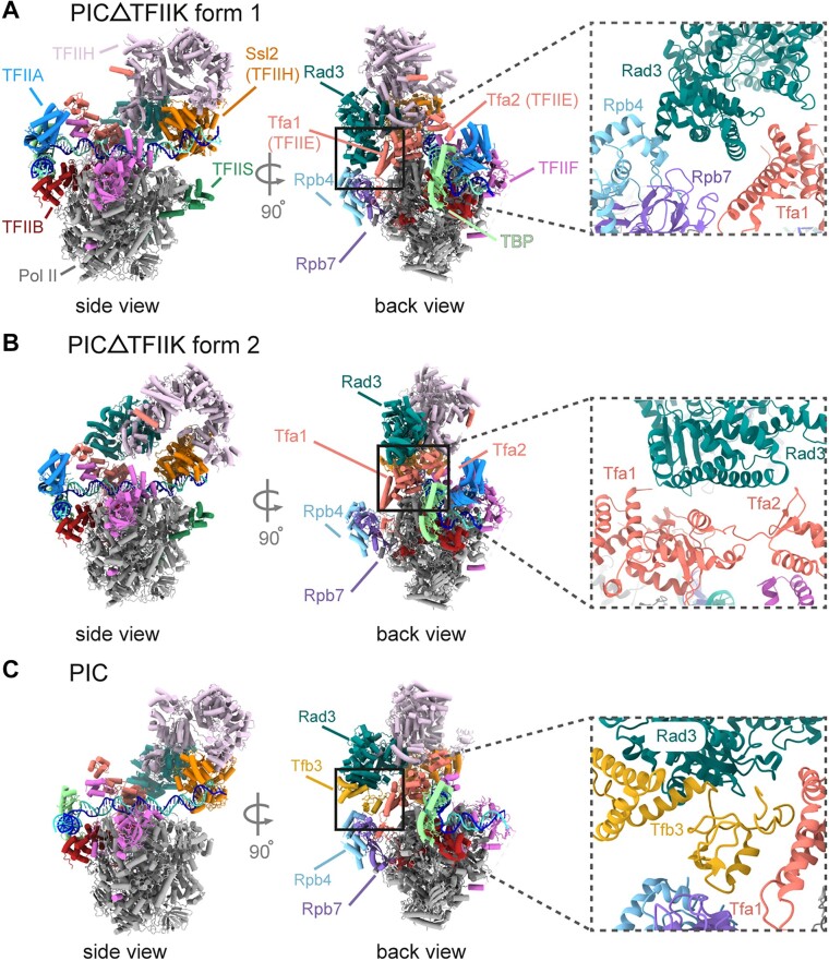
Figure 4.
Cryo-EM structures of PIC-ΔTFIIK. (A) Cryo-EM map of form 1 of PIC-ΔTFIIK in side (left) and back (right) views with a corresponding model in the lower panel. Rad3 (sea green) is in direct contact with Rpb4 (light blue) and Tfa1 (salmon). (B) Same as panel (A), but for form 2. (C) Structure of the PIC containing TFIIK (PDB ID: 7ML0) is shown as cartoon in the same orientations as in panels (A) and (B).

