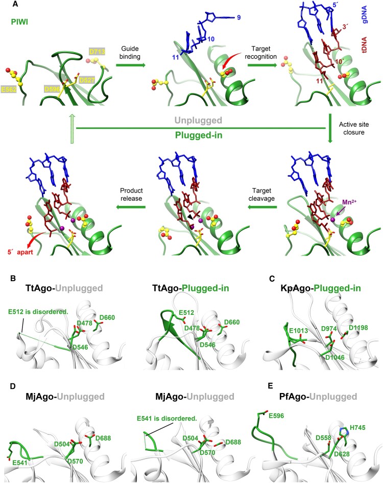
Figure 4.
Comparison of the catalytic tetrad in KmAgo with other species. (A) The conformational changes within the active site of KmAgo during target recognition and catalysis. The residues located within the active site are highlighted in yellow, with E562 and D713 represented as ball-and-stick models. In the initial stages of guide binding and target recognition, the active site is unplugged (upper row). This is followed by the ‘plugged-in’ of the glutamate finger, binding of the catalytic metal, and activation of catalysis (lower row). The accompanying variations in D713 and product release are denoted by red arrows. The PDB accession numbers are (from the upper left corner, clockwise): 8XHS, 8XHV, 8XJX, 8XK0, 8XK3, 8XK4. (B–E) States whose conformations can be deduced from the other known structures are marked as green (plugged-in) and gray (unplugged). (B) TtAgo (left panel, 3DLH; right panel, 4NCB). (C) KpAgo (4F1N). (D) MjAgo (left panel, 5G5S; right panel, 5G5T). (E) PfAgo (1U04).

