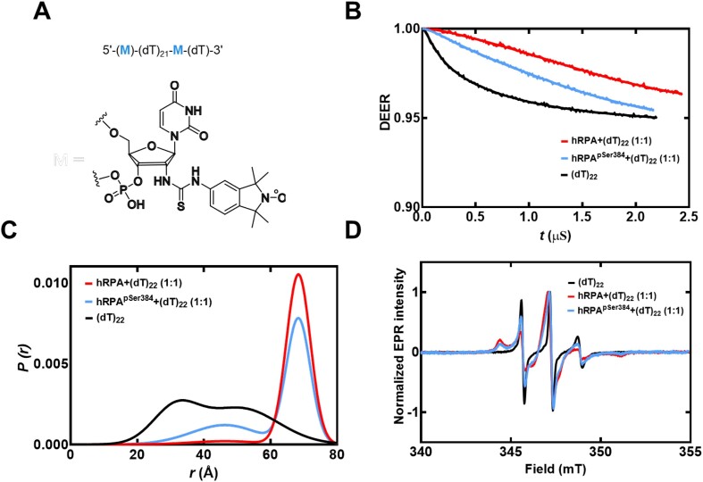
Figure 5.
DEER spectroscopy of RPA and RPA-pSer384 bound to ssDNA. (A) Structure of the isoindoline nitroxide spin label and the position on the (dT) oligonucleotide. (B) Raw DEER decays and fits are presented for the experimentally determined distance distributions P(r) (C). (D) CW EPR spectra of labeled ssDNA in the absence and presence of RPA and RPA-pSer384.

