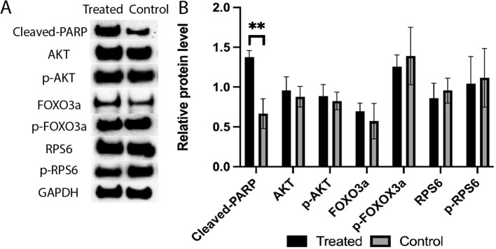
Figure 9.
(A) Expression levels of cleaved-PARP, AKT, p-AKT, FOXO3a, p-FOXO3a, RPS6, and p-RPS6 proteins in the 4-HC-treated and control groups, detected by Western blotting. GAPDH was used as an internal control. (B) Protein expression levels were quantified using Image J, data mean ± SD, **P < 0.01.

