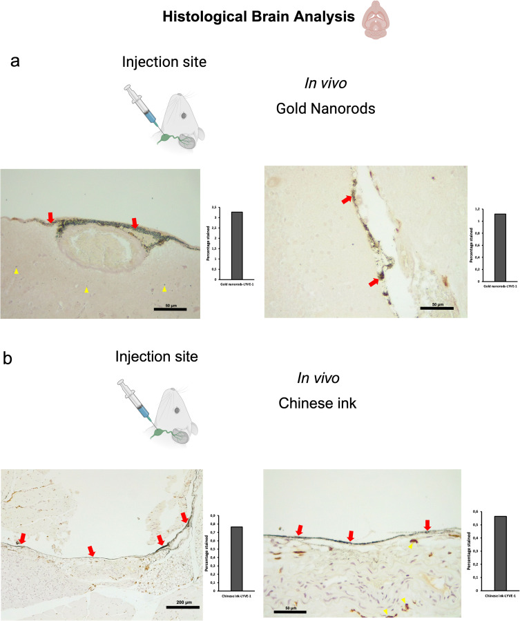
Figure 5.
Retrograde directional flow analysis by brain histology after in vivo nanoparticle administration into the deep cervical lymph node. (a) Combined Gold Enhancement and anti-LYVE-1 immunohistochemistry showed gold nanorods within meningeal lymphatic vessels (red arrows) and the brain parenchyma (yellow arrows), with no staining within cerebral arteries (n=3). (b) Meningeal lymphatic vessels stained with anti-LYVE-1 immunohistochemistry and colocalized with Chinese ink nanoparticles (red arrows). Chinese ink was also identified in the brain parenchyma (yellow arrows) (n=4).
Abbreviation: LYVE-1, lymphatic vessel endothelial hyaluronan receptor-1.

